IT Band Syndrome, also called Iliotibial Band Syndrome, is a frequent overuse injury in athletes, especially runners and cyclists. This condition arises when the iliotibial band, a thick tissue band running along the outside of the thigh from the hip to the knee, becomes tight or inflamed. It can lead to considerable pain and impede performance.
Effective treatment is essential for recovery and preventing further injury. In the first instance, physiotherapy is the mainstay of treatment.
The treatment will involve:
- Rest, Ice, stretching exercises
- Specific exercises targeting the muscles supporting the hip and knee in particular
- Adjusting training load
- Taping of the knee
- Biomechanical assessment of running technique and modification
- Soft tissue manipulation
- Nonsteroidal anti-inflammatory drugs (NSAIDs)
If symptoms are persistent or severe and physiotherapy is not making any impact on symptoms, then injection therapy is the next step to deal with this condition effectively. Sonoscope offers pinpoint accurate ultrasound-guided injections directly targeting the structure affected.
How Is IT Band Syndrome Diagnosed?
How is IT Band Syndrome diagnosed? This is an important question, especially if you are experiencing pain on the outside of your knee. IT Band Syndrome, or Iliotibial Band Syndrome, is a common injury among runners and other athletes. The iliotibial band is a thick band of tissue that runs along the outside of your thigh, from your hip to your knee. When this band becomes tight or inflamed, it can cause pain and discomfort, especially during physical activities.
The first step in diagnosing IT Band Syndrome is usually a visit to a doctor or a physical therapist. They will start by asking you about your symptoms and your medical history. They will want to know when the pain started, where exactly it hurts, and what activities seem to make it worse. They will also ask about your exercise routine and any recent changes in your activity level.
Next, the doctor will perform a physical examination. They will look at your leg and knee, checking for areas of tenderness and swelling. They may press on the outside of your knee to see if it is painful. They will also observe your posture and gait to see if there are any abnormalities that could be contributing to your symptoms.
In addition to the physical examination, the doctor may ask you to perform certain movements or exercises. These tests are designed to put stress on the iliotibial band and see if they reproduce your symptoms. For example, they might ask you to bend and straighten your knee while pressing on the outside of it. If this causes pain, it could be a sign of IT Band Syndrome.
Sometimes, imaging tests may be used to help diagnose IT Band Syndrome. An X-ray can rule out other conditions that might be causing your knee pain, such as a fracture or osteoarthritis. An MRI (Magnetic Resonance Imaging) scan can provide a detailed image of the soft tissues in your leg, including the iliotibial band. This can help to confirm the diagnosis and rule out other possible causes of your symptoms.
However, imaging is not always necessary for diagnosing IT Band Syndrome. In many cases, a thorough history and physical examination is enough to make the diagnosis. The doctor or physical therapist will then discuss the findings with you and explain the next steps for treatment.
In summary, diagnosing IT Band Syndrome involves a combination of taking a medical history, performing a physical examination, and sometimes using imaging tests. The key to successful treatment and prevention is to address the underlying causes and make necessary changes to your activity routine. If you think you might have IT Band Syndrome, it is important to see a doctor or physical therapist who can help you develop a plan to manage your symptoms and prevent future problems.
Diagnostic Ultrasound:
Diagnostic ultrasound is very helpful for assessment of iliotibial band friction syndrome. As this is a dynamic modality we we will be able to see the IT band as it moves over the lateral epicondyle of the knee during extension and flexion of the knee. This allows us to evaluate for any thickening of the ITB but also to identify any iliotibial bursitis which is indicated by fluid in the iliotibial bursa.
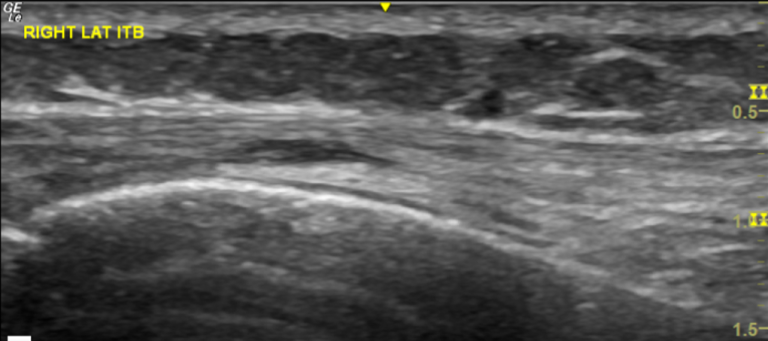
ruling out other potential sources of lateral knee pain such as ligamentous injuries or meniscal tears.
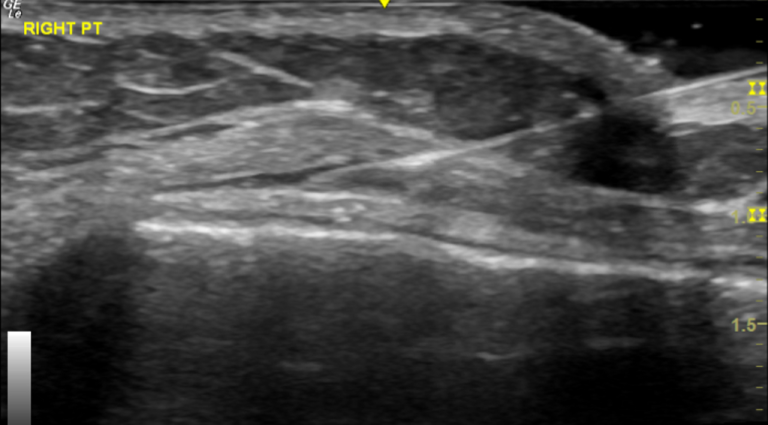
The ultrasound-guided injection procedure allows very specific targeting of the iliotibial bursa. The bursa is clearly identifiable on ultrasound and for a skilled practitioner the needle is then accurately guided in the ITB bursa which lies superficial of the lateral femoral condyle and underneath the iliotibial band. The accurate administration of steroid also avoids potential weakening of the IT band by inadvertent injection in the body of the IT band which is very realistic possibility if this injection is done unguided.
Conclusion:
ITB friction syndrome is a frequent overuse injury which often affects runners but also cyclists. This is a condition that can lead to significant pain and loss of function making it difficult if not impossible to exercise. Initial management is with non-invasive options such as physiotherapy treatment rest, stretches, strengthening exercises and nonsteroidal anti-inflammatory drugs. If these options are not sufficient then ultrasound-guided steroid injection is a very effective option to quickly reduce inflammation and thus reduce pain.
Diagnostic ultrasound is very helpful in accurately diagnosing the condition and then also allowing accurate injection of a steroid in the inflamed bursa between IT band and lateral femoral condyle
In the Sonoscope clinic we provide a One-Stop-Clinic which includes clinical diagnosis, diagnostic ultrasound and ultrasound-guided injection all within the One session.